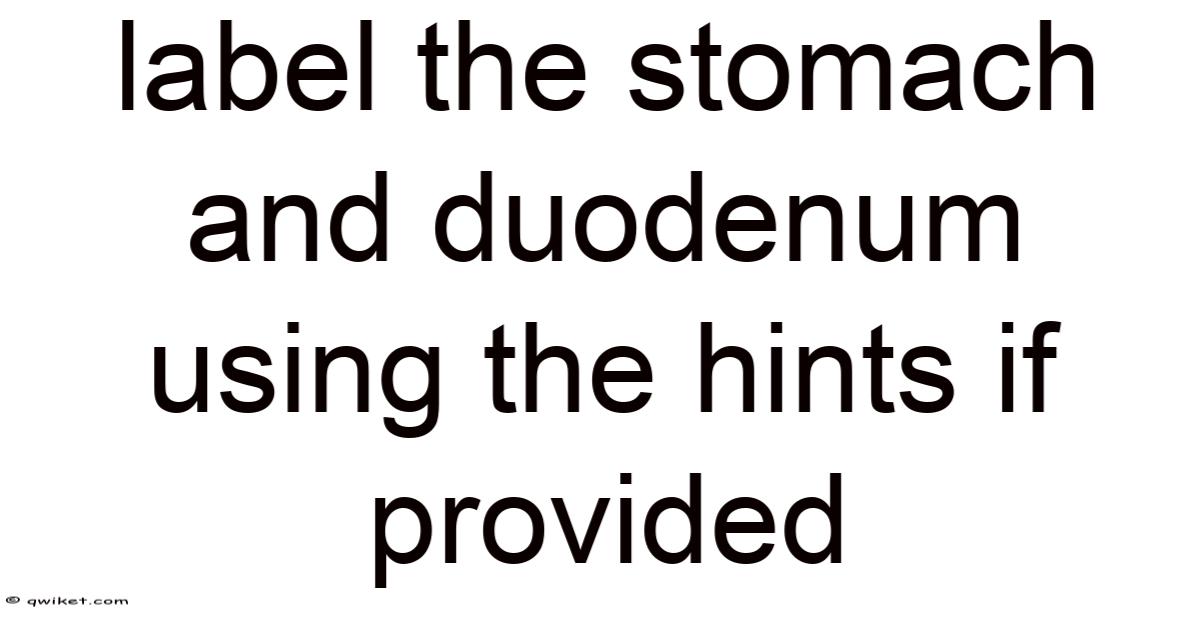
Labeling the stomach and duodenum using the hints if provided is a useful anatomy exercise because it helps students connect structure names with real positions in the digestive system. The stomach is the J-shaped organ that receives food from the esophagus, mixes it with gastric juices, and slowly releases it into the du
Following the identification of the stomach and duodenum, it becomes evident the precise articulations within the gastrointestinal tract play a critical role in digestion and nutrient absorption. The stomach, known for its powerful muscular contractions, acts as a central hub where food is broken down and mixed with gastric secretions, preparing the substrate for the subsequent process in the small intestine. Concurrently, the duodenum, the first segment of the small intestine, is intricately positioned at the junction of the stomach and the jejunum, where its brief role in initial digestion and absorption is complemented by its close proximity to the duodenal vein and lymphatic system, enhancing its functional efficiency. These structural landmarks not only guide anatomical understanding but also influence the physiological behaviors of digestion and the passage of food through the system, underscoring the complexity and interdependence inherent in the digestive process. Their precise placement and function thus serve as a foundation for the seamless operation of the entire gastrointestinal tract, highlighting the importance of correct anatomical mapping in medical education and clinical practice. A thorough grasp of these concepts is essential for accurately diagnosing and treating gastrointestinal disorders, emphasizing the critical link between anatomy and clinical physiology. At the end of the day, the study of the stomach and duodenum, along with their respective roles within the digestive pathway, encapsulates a fundamental aspect of human health and nutrition, reinforcing the necessity of a comprehensive understanding of the body's nuanced systems.
No fluff here — just what actually works.
The integration of anatomical knowledge with physiological processes becomes even more apparent when considering the collaborative roles of accessory organs like the liver, gallbladder, and pancreas in duodenal function. The duodenum receives bile from the liver and gallbladder, which emulsifies fats, and pancreatic juice rich in digestive enzymes, a process regulated by hormones such as secretin and cholecystokinin. Worth adding: these secretions, triggered by the acidic and fatty chyme entering the duodenum, neutralize gastric acidity and initiate enzymatic breakdown of nutrients, illustrating the organ’s role as a critical transition zone. Meanwhile, the stomach’s muscular layers—the longitudinal, circular, and oblique—coordinate peristaltic waves to churn and propel contents, aided by the pyloric sphincter, which regulates emptying into the duodenum Simple, but easy to overlook. And it works..
Clinically, understanding these structures is vital for addressing disorders such as peptic ulcers, which often originate in the stomach or duodenal lining due to Helicobacter pylori infection or excessive acid exposure. Because of that, conditions like celiac disease, which damages intestinal villi, can impair duodenal absorption, leading to malnutrition. Additionally, procedures such as endoscopy rely on precise anatomical landmarks to visualize and treat lesions, while surgeries like gastrojejunostomy bypass the duodenum in cases of obstruction or disease.
This interplay between structure and function underscores the necessity of mastering anatomical details for effective clinical reasoning. So by appreciating how the stomach and duodenum collaborate with neighboring organs and systems, healthcare professionals can better diagnose, manage, and innovate treatments for digestive ailments. At the end of the day, the synergy of anatomical precision and physiological insight remains indispensable in advancing both medical education and patient care, ensuring that foundational knowledge translates into practical, life-saving interventions.
This is where a lot of people lose the thread.